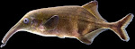
The transparency of zebrafish larvae makes the animals ideal models for seeing deep into tissues. CHARLES MAZEL/VISUALS UNLIMITED/CORBIS
Tiny fish trapped in a virtual world provide a window into complex brain connections.
Virginia Hughes
A recently hatched zebrafish is swimming upriver for the first time. Its big round eyes, bulging on the front of its eyelash-sized body, scan the surroundings. Suddenly, it sees the scenery flying forwards as a gentle current pushes it backwards. The fish flicks its tail to try to stay in place. Or so it thinks.
In reality, the baby fish is paralysed and suspended in a water-filled Petri dish by glass pipettes. The dish sits on the stage of a US$100,000 microscope in the corner of a darkened, cluttered laboratory. A film, projected from below, has transported the fish to a virtual world in which moving bands of light and dark simulate passing underwater scenery.
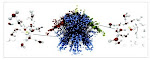
Although the fish doesn't move, the motor neurons that control its tail are firing away, just as if it were swimming. And when fed into a computer, those signals can control the video display, giving the fish nearly every sign that it is swimming normally. All the while, Florian Engert's microscope peers deep into the fish's tiny, translucent brain to watch neurons glow green as they fire (see ‘A river of deceit’).
Virginia Hughes
A recently hatched zebrafish is swimming upriver for the first time. Its big round eyes, bulging on the front of its eyelash-sized body, scan the surroundings. Suddenly, it sees the scenery flying forwards as a gentle current pushes it backwards. The fish flicks its tail to try to stay in place. Or so it thinks.
In reality, the baby fish is paralysed and suspended in a water-filled Petri dish by glass pipettes. The dish sits on the stage of a US$100,000 microscope in the corner of a darkened, cluttered laboratory. A film, projected from below, has transported the fish to a virtual world in which moving bands of light and dark simulate passing underwater scenery.
Although the fish doesn't move, the motor neurons that control its tail are firing away, just as if it were swimming. And when fed into a computer, those signals can control the video display, giving the fish nearly every sign that it is swimming normally. All the while, Florian Engert's microscope peers deep into the fish's tiny, translucent brain to watch neurons glow green as they fire (see ‘A river of deceit’).
Nature 493, 466–468 (24 January 2013)